
Yesterday as I was listening to a talk about diffusion tensor imaging, a professor asked about the influences of head motion on DTI data, and whether it could lead to spurious effects. Another professor vigorously denied it, stating that it was much more of a problem for bread and butter FMRI analyses, and in particular resting state functional connectivity analyses. At one point he became so animated that his monocle fell off, his collar stud came undone, and eventually he had to be physically restrained by the people sitting next to him. It was then that I knew that I should pay heed, for it is unlikely that a scientist becomes highly excited and talkative over matters that are trivial; in short, I could sense that he was talking about something important.
I have done few functional connectivity analyses in my brief life, but what I understand about them is this: You take the timecourse of one voxel - or the average timecourse over a group of voxels, also known as a "seed" - and then compare that timecourse with the timecourse of every other voxel in the brain. (When I speak of timecourses, I mean the sampled signal over time.) If it is a good fit - in other words, if there is a significantly high correlation between the timecourses - then we say that the two regions are functionally connected. This is a bit of a misnomer, as we cannot make any direct inferences about any "real" connectivity from two correlated timecourses; but it can serve as a good starting point for more sophisticated analyses, such as psychophysiological interactions (PPI; also known as context-dependent correlations) which measure changes in functional connectivity as a function of task context. For example: Does the timecourse correlation between cognitive control regions and reward-related areas change depending on whether the subject is instructed to upregulate or downregulate their gut reactions to rewarding stimuli?
One of the most popular variations of functional connectivity is something called resting state functional connectivity (rsFC), where a subject is simply instructed to relax and listen to Barry Manilow* while undergoing scanning. Functional connectivity maps are then calculated, and usually a comparison is made between a control group and an experimental or patient group, such as schizophrenics. For us FMRI researchers, this is about as close as we can get to simulating a person's natural environment where they would be relaxing and thinking about nothing in particular; except that they're in an extremely tight-fitting, noisy tube, and unable to move in any direction more than a few millimeters. Other than that, though, it's pretty normal.
These types of experiments have become all the rage in recent years, with several studies claiming to have found meaningful resting-state differences between healthy controls and several different patients populations such as schizophrenics, depressives, Nickelback fans, and drug addicts. However, a few publications have called into question some of these results, stating that many of these differences could be due to head motion. As we've talked about before, head motion can be a particularly insidious confound in any experiment, but it is especially troublesome for functional connectivity analyses. This can arise due to systematic differences between control and patient populations that are possibly confounded with motion. Take, for example, an experiment contrasting young versus older populations. Older populations are known to move more, and any observed differences in functional connectivity may be due solely to this increased motion, not underlying neural hemodynamics.
A study by Van Dijk, Sabuncu, & Bruckner (2012) looked at this in detail by scanning over a thousand (!) subjects, and binning them into ten groups based on increasing amounts of motion (e.g., group 1 had the least amount of motion, while group 10 had the most motion). The authors found decreased functional connectivity in the "default network" of the brain - usually referring to the functional connectivity between the medial prefrontal cortex and retrosplenial cingulate cortex -, decreased connectivity in the frontal-parietal network, and slightly increased local connectivity among clustered voxels, simply based on motion alone. (Keep in mind that each bin of subjects were matched as closely as possible on all other demographic measures.) Furthermore, even when comparing bins of subjects closely matched for motion (e.g., bins 5 and 6), small but significant differences in functional connectivity were seen.
Lastly, a subset of subjects were rescanned in order to see whether motion was reliable; in other words, if a subject that moved a large amount on one day had the same tendency to move a large amount on the next day. A clear correlation was found between scanning subjects, suggesting that motion might need to be treated as a trait or individual difference, just like any other.
So, what to do? A few recommendations are to match subjects for motion, correct motion prospectively (Ward et al, 2000), and regress out motion when performing a group-level analysis, as you would any other covariate. Apparently traditional methods of motion correction on a subject-by-subject basis are not enough, and increasing awareness of the pitfalls of between-subject motion is important for evaluating current functional connectivity analyses, and for conducting your own experiments.
This study hit me in the face like a wet mackerel since I am beginning to investigate a recent AFNI tool, 3dReHo, to do local functional connectivity analyses for publicly available datasets on the ABIDE website. However, as far as I can tell, motion limits were not used as exclusionary criteria, which may be a possible confound when examining, say, autistic children to controls. More to come soon. Or not.
*I Don't Want to Walk Without You
I have done few functional connectivity analyses in my brief life, but what I understand about them is this: You take the timecourse of one voxel - or the average timecourse over a group of voxels, also known as a "seed" - and then compare that timecourse with the timecourse of every other voxel in the brain. (When I speak of timecourses, I mean the sampled signal over time.) If it is a good fit - in other words, if there is a significantly high correlation between the timecourses - then we say that the two regions are functionally connected. This is a bit of a misnomer, as we cannot make any direct inferences about any "real" connectivity from two correlated timecourses; but it can serve as a good starting point for more sophisticated analyses, such as psychophysiological interactions (PPI; also known as context-dependent correlations) which measure changes in functional connectivity as a function of task context. For example: Does the timecourse correlation between cognitive control regions and reward-related areas change depending on whether the subject is instructed to upregulate or downregulate their gut reactions to rewarding stimuli?
One of the most popular variations of functional connectivity is something called resting state functional connectivity (rsFC), where a subject is simply instructed to relax and listen to Barry Manilow* while undergoing scanning. Functional connectivity maps are then calculated, and usually a comparison is made between a control group and an experimental or patient group, such as schizophrenics. For us FMRI researchers, this is about as close as we can get to simulating a person's natural environment where they would be relaxing and thinking about nothing in particular; except that they're in an extremely tight-fitting, noisy tube, and unable to move in any direction more than a few millimeters. Other than that, though, it's pretty normal.
These types of experiments have become all the rage in recent years, with several studies claiming to have found meaningful resting-state differences between healthy controls and several different patients populations such as schizophrenics, depressives, Nickelback fans, and drug addicts. However, a few publications have called into question some of these results, stating that many of these differences could be due to head motion. As we've talked about before, head motion can be a particularly insidious confound in any experiment, but it is especially troublesome for functional connectivity analyses. This can arise due to systematic differences between control and patient populations that are possibly confounded with motion. Take, for example, an experiment contrasting young versus older populations. Older populations are known to move more, and any observed differences in functional connectivity may be due solely to this increased motion, not underlying neural hemodynamics.
A study by Van Dijk, Sabuncu, & Bruckner (2012) looked at this in detail by scanning over a thousand (!) subjects, and binning them into ten groups based on increasing amounts of motion (e.g., group 1 had the least amount of motion, while group 10 had the most motion). The authors found decreased functional connectivity in the "default network" of the brain - usually referring to the functional connectivity between the medial prefrontal cortex and retrosplenial cingulate cortex -, decreased connectivity in the frontal-parietal network, and slightly increased local connectivity among clustered voxels, simply based on motion alone. (Keep in mind that each bin of subjects were matched as closely as possible on all other demographic measures.) Furthermore, even when comparing bins of subjects closely matched for motion (e.g., bins 5 and 6), small but significant differences in functional connectivity were seen.
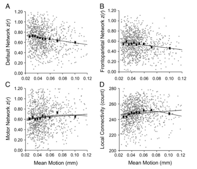 |
| Figure 3 from Van Dijk et al (2012). Functional connectivity among different networks measured as a function of head motion. Both linear and nonlinear (quadratic) terms were modeled to fit the data. |
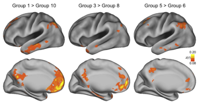 |
| Figure 4 from Van Dijk et al (2012). Note the comparison on the far right between groups 5 and 6; the mean motion difference between these two groups is a few thousandths of a millimeter, but noticeable functional connectivity differences are still seen between the two groups. |
Lastly, a subset of subjects were rescanned in order to see whether motion was reliable; in other words, if a subject that moved a large amount on one day had the same tendency to move a large amount on the next day. A clear correlation was found between scanning subjects, suggesting that motion might need to be treated as a trait or individual difference, just like any other.
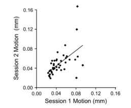 |
| Figure 5 from Van Dijk et al (2012). There is a robust correlation between the movement of scanning sessions, even with the outliers removed (marked in diamonds). |
So, what to do? A few recommendations are to match subjects for motion, correct motion prospectively (Ward et al, 2000), and regress out motion when performing a group-level analysis, as you would any other covariate. Apparently traditional methods of motion correction on a subject-by-subject basis are not enough, and increasing awareness of the pitfalls of between-subject motion is important for evaluating current functional connectivity analyses, and for conducting your own experiments.
This study hit me in the face like a wet mackerel since I am beginning to investigate a recent AFNI tool, 3dReHo, to do local functional connectivity analyses for publicly available datasets on the ABIDE website. However, as far as I can tell, motion limits were not used as exclusionary criteria, which may be a possible confound when examining, say, autistic children to controls. More to come soon. Or not.
*I Don't Want to Walk Without You