Running Current Through Your Brain Improves Performance, Not As Likely To Kill You As You Think

For many people placing a nine-volt battery on their tongue, like sex, can be a fun, exciting activity, sometimes resulting in death. Based on this observation, Reinhart and Woodman (2014) decided to turn the brain into an improvised battery by placing electrodes on different areas of the skull and zapping it with enough current to toast a frozen hotpocket. If this sounds insane to you, then you are obviously not a cognitive neuroscientist - as I have said before, we live for these kinds of experiments.

However, the researchers had good reasons for doing this. First of all, they are scientists, and they did not spend nine years on their doctorate only to justify themselves to the likes of you. Second, directly messing with brain activity can lead to valuable scientific insights, such as how much you have to pay an undergraduate to have them consent to turn their brain into a microwave oven. But third, and most important, delivering direct current through a pair of electrodes can increase or decrease certain patterns of neural activity - specifically, the error-related negativity (ERN) following an error trial.

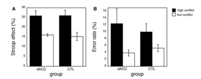
The ERN is a negative deflection in voltage over the medial frontal lobes that correlates with behavior adjustment and error correction in the future. For example, if I commit what some narrow-minded, parochial individuals consider an error, such as asking out my girlfriend's sister, the larger my ERN is, the less likely I am to make that same mistake in the future. Similarly, with experiments such as the Stroop task, or any performance task, the larger the ERN after committing an error, the greater the probability of making a correct response on the next trial. Furthermore, whereas the ERN usually occurs immediately after the response is made, another related signal, the feedback related negativity (FRN) occurs once feedback is received. In sum, larger ERNs and FRNs generally lead to better future performance.

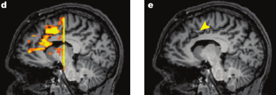
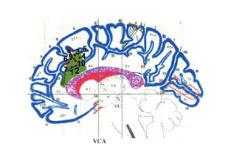
This is exactly what the experimenters manipulated when they sent current through the medial frontal area of the brain, corresponding to the dorsal anterior cingulate cortex (dACC) and supplementary motor area (SMA) cortical regions. The electrode over this area was changed to either a cathode (i.e., positively charged, or where the electrons flowed toward) or an anode (i.e., negatively charged, or where the electrons flowed away from). If the electrode was a cathode, the ERN decreased significantly, whereas if the electrode was an anode, the ERN significantly increased.

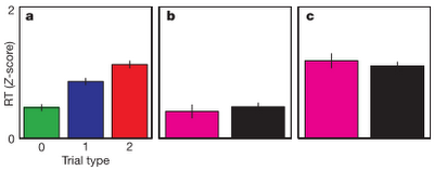
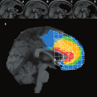
Figure 1 from Reinhart & Woodman (2014). Panels A and D represent the current distribution throughout the medial prefrontal cortex. B: Stop-signal task used in the experiment. A stop signal leads to a greater chance of screwing up, and the longer the delay between the cue and the stop signal, the more difficult it is to stop a response. This is what physicists and individuals with severe incontinence refer to as an "event horizon." C: Placement of Cathode or Anode on the medial frontal surface, along with a sham condition. Lower panel: Difference in ERN and FRN dependent on whether the fronto-medial electrode is an Anode or Cathode.

As interesting as these neural differences are, however, the real punch of the paper lies in the behavioral changes. Participants who had an anode placed over their cingulate and SMA areas not only showed greater ERN and FRN profiles, but also steep gains in their accuracy and improvements in reaction time. For regular trials which did not include a distracting stop signal, anode subjects were markedly faster than in the cathode and sham conditions, and in both regular and stop-signal trials, accuracy nearly reached a hundred percent.


Nor were these gains limited to the duration of the experiment; in fact, behavioral improvements could last as long as five hours after switching on the current. These results make for wild and reckless speculations about what could be done with this kind of setup; one could imagine creating caps for students which get them "juiced up" for exams, hats for the elderly to help them find their Mysteriously Disappearing Reading Glasses, or modified helmets for soldiers which allow them get even better at BSU (blowing stuff up). Because, after all, what's the use of a scientific result if you can't weaponize it?

More figures and results from experiments further extending and confirming their results can be seen in the paper, found here.

Lesion Studies: Recent Trends

A couple weeks ago I blogged hard about a problem presented by lesion studies of the anterior cingulate cortex (ACC), a broad swath of cortex shown to be involved in aspects of cognitive control such as conflict monitoring (Botvinick et al, 2001) and predicting error likelihood (Brown & Braver, 2005). Put simply: The annihilation of this region, either through strokes, infarctions, bullet wounds, or other cerebral insults, does not result in a deficit of cognitive control, as measured through reaction time (RT) in response to a variety of tasks, such as Stroop tasks - a task that requires overriding prepotent responses to a word presented on a screen, as opposed to the color of the ink that the word is written in - and paradigms which involve task-switching.

In particular, a lesion study by Fellows & Farah (2005) did not find a significant RT interaction of group (either controls or lesion patients) by condition (either low or high conflict in a Stroop task; i.e., either the word and ink color matched or did not match), suggesting that the performance of the lesion patients was essentially the same as the performance of controls. This in turn prompted the question of whether the ACC was really necessary for cognitive control, since those without it seemed to do just fine, and were about a pound lighter to boot. (Rimshot)

However, a recent study by Sheth et al (2012) in Nature examined six lesion patients undergoing cingulotomy, a surgical procedure which removes a localized portion of the dorsal anterior cingulate (dACC) in order to alleviate severe obsessive-compulsive symptoms, such as the desire to compulsively check the amount of hits your blog gets every hour. Before the cingulotomy, the patients performed a multisource interference task designed to elicit cognitive control mechanisms associated with dACC activation. The resulting cingulotomy overlapped with the peak dACC activation observed in response to high-conflict as contrasted with low-conflict trials (Figure 1).

Figure 1 reproduced from Sheth et al (2012). d) dACC activation in response to conflict. e) arrow pointing to lesion site

Furthermore, the pattern of RTs before surgery followed a typical response pattern replicated over several studies using this task: RTs were faster for trials immediately following trials of a similar type - such as congruent trials following congruent trials, or incongruent trials following incongruent trials - and RTs were slower for trials which immediately followed trials of a different type, a pattern known as the Gratton effect.

The authors found that global error rates and RTs were similar before and after the surgery, dovetailing with the results reported by Fellows & Farah (2005); however, the modulation of RT based on previous trial congruency or incongruency was abolished. These results suggest that the ACC functions as a continuous updating mechanism modulating responses based on the weighted past and on trial-by-trial cognitive demands, which fits into the framework posited by Dosenbach (2007, 2008) that outlines the ACC as part of a rapid-updating cingulo-opercular network necessary for quick and flexible changes in performance based on task demands and performance history.

a) Pre-surgical RTs in response to trials of increasing conflict. b, c) Post-surgical RTs showing no difference between low-conflict trials preceded by either similar or different trial types (b), and no RT difference between high-conflict trials preceded by either similar or different trial types (c).


Above all, this experiment illustrates how lesion studies ought to be conducted. First, the authors identified a small population of subjects about to undergo a localized surgical procedure to lesion a specific area of the brain known to be involved in cognitive control; the same subjects were tested before the surgery using fMRI and during surgery using single-cell recordings; and interactions were tested which had been overlooked by previous lesion studies. It is an elegant and simple design; although I imagine that testing subjects while they had their skulls split open and electrodes jammed into their brains was disgusting. The things that these sickos will do for high-profile papers.

(This study may be profitably read in conjunction with a recent meta-analysis of lesion subjects (Gläscher et al, 2012; PNAS) dissociating cortical structures involved in cognitive control as opposed to decision-making and evaluation tasks. I recommend giving both of these studies a read.)

Lesion Studies: Thoughts

(Note: I recently completed my candidacy exam, which involved writing a trio of papers focusing on different aspects of my research. Most of this post is cannibalized from a section I wrote on lesion studies of the anterior cingulate cortex, which produce counterintuitive results when contrasted to lesions of other areas, such as the DLPFC and OFC, which do indeed seem to disrupt the processes that those regions are implicated in from the neuroimaging literature.

My work primarily involves healthy people with intact brains, and observing indirect measures of neural firing through tracking slow blood flow changes in the brain. However, "activation" as defined by fMRI is not the same as the underlying neural dynamics, and, barring invasive single-cell recordings, we have few options for directly measuring neural firing in response to different tasks and psychological contexts. This caveat inherent in fMRI research becomes particularly important when interpreting the results of lesion studies.) 

Although the majority of the neuroimaging literature has implicated the dACC as playing a critical role in the signaling for cognitive control when necessary, the most direct test of a brain structure’s necessity in a cognitive process is through examining subjects presenting with lesions in that part of the brain. For example, if it can be demonstrated that a subject without an ACC still performs equivalent to controls on tasks involving cognitive control, then that would argue against the necessity of that area’s involvement in the hypothesized cognitive process. Studies involving human subjects with lesions are relatively rare and suffer from low power, but can still reveal important aspects of neural functioning.


The ACC, in particular, has been the subject of several lesion studies that have shown conflicting and counterintuitive results. For example, a single-subject lesion study of a patient with left ACC damage exhibited both smaller ERNs and increased RT in response to incongruent stimuli in a spatial Stroop paradigm. This study showed that conflict monitoring and error detection, at least in this patient, do not both come from the same area of ACC, suggesting that these processes occur in different areas. However, while the ERN was shown to be attenuated in the patient, the conflict response (a waveform called the N450) was actually enhanced (Swick & Turken, 2002). This suggests that conflict monitoring occurs in a nearby prefrontal area, such as the DLPFC, before information about the conflict is sent to the ACC.


Figure of the lesion for the single subject analyzed by Turken & Swick (2002). Overlaid are coordinates of peak activation for conflict-related tasks from other studies.


On the other hand, a lesion study conducted by Fellows & Farah (2005) compared the performance of individuals with dACC lesions to that of controls across a battery of tasks hypothesized to involve cognitive control. These tasks included a Stroop task and a go-nogo task which are known to elicit significantly greater increases in RT after errors, and to induce significantly greater amounts of errors during incongruent trials. The results showed no significant interactions between group and task, suggesting that the dACC is not necessary for the implementation of cognitive control. Furthermore, the authors pointed out that tasks involving cognitive control may be confounded with emotional responding, which in turn could simply be associated with the ACC's involvement in regulating muscle tone. In any case, it is apparent that although this structure is somehow associated with cognitive control, it is not strictly necessary for it. 


Figure showing group overlap of lesions in the Fellows & Farah (2005) study.  Circles and squares represent an overlay of a meta-analysis by Bush et al (2000), with circles representing peak activations for cognitive tasks, and squares representing peak activations for emotional tasks.

Comparison of Stroop effect (measured in percent signal change from mean congruent trial RT) and error rate between lesion patients and controls. No significant difference was found on either measure between the two groups.

In sum, these lesion studies suggest that the dACC may not be indispensable for signaling the DLPFC to implement cognitive control. However an alternative explanation is that patients with ACC lesions are usually ipsilateral, and that furthermore they may be compensating for required cognitive control by recruiting nearby cortical areas. However, two lines of evidence argue against this interpretation. First, one of the lesion patients examined in the Fellows & Farah (2005) had extensive medial ACC damage encompassing dACC bilaterally, but showed a similar pattern of error rates and RT difference between congruent and incongruent conditions as did the other lesion patients and the control group. Secondly, lesion studies of other areas of the brain – such as the orbitofrontal cortex – have shown that those regions appear to be specific to the cognitive processes they are hypothesized to be involved in. For example, patients with OFC lesions exhibit significantly impaired performance in decision-making tasks such as the Iowa Gambling Task and Wisconsin Card Sorting Task, as well as decreased autonomic activity in response to highly risky gambles (Bechara et al, 1994). Even though the patients in this study had suffered from their lesions for a comparable amount of time as the lesion subjects in the Fellows & Farah (2005) study, there was no evidence of recruitment of other cortical areas in order to support their deficits in decision-making.

However, although these lesion studies have shown no significant differences in error rates between the lesion patients and controls, other experiments have revealed that patients with ACC damage are less likely to correct for their mistakes on trials immediately following an error. In addition, patients with ACC lesions are less likely to be aware that an error has occurred (Swick & Turken, 2002). These results suggest that there may be a necessary role for of the ACC for the actual detection of errors, which would be consistent with the hypothesis that this area is involved in the comparison of actions against their predicted outcomes. How lesions affect the transfer of information from the ACC to the DLPFC and other cortical regions supposedly involved in the implementation of cognitive control, however, is less well understood.

Bottom line: If the inferences from neuroimaging studies are to believed, then the ACC is necessary somehow for cognitive control or executive function; however, lesion studies belie this claim, suggesting perhaps that the necessary processes for these cognitive functions take place elsewhere and merely light up the ACC as some sort of epiphenomenon. Admittedly, I am unsure of what to make of all this. The most useful experiments to carry out, in my opinion, would be to apply transcranial magnetic stimulation (TMS) to temporarily knock out this area in healthy controls, and then observe what happens; however, as TMS is only able to disrupt neural firing on surface areas of the cortex, stimulation of deeper areas remains impractical. With continuing advances in the ability of TMS to stimulate deeper cortical (and, possibly, subcortical?) structures, we may get a better grasp of what is going on.